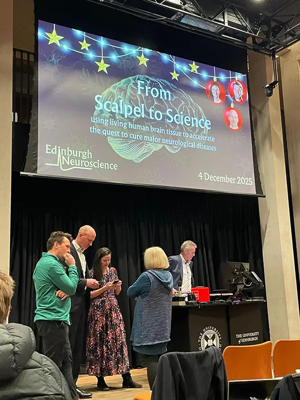
An Evening with Edinburgh Neuroscience
Don’t you love when an idea is so simple and obviously a good one. Such as conducting neuroscience research on living, human brain tissue. Of course the logistics involved in transforming that from an idea into a lab are vastly complicated. However, it had become a reality at the University of Edinburgh.
Last week I had the pleasure of swapping my desk for a lecture organised by Edinburgh Neuroscience to hear how this is being achieved. It was a trip down memory lane back to my uni days. But the night was really about astonishing, ingenious, and pragmatic new science.
What models are currently being used for neuroscience research?
Historically, conclusions have been drawn from testing on drosophila, mice or large animals. You don’t need to be a neuroscientist to imagine that these aren’t a direct parallel for what might be happening in human brains. While these models are essential they have limits.
Indeed, two recent research papers from the Living Brain Project in Mount Sinai have shown that even dead, human brain tissue has different gene1 and protein expression2 compared with living human brain tissue.
So it was very exciting to hear how this cross functional team in Edinburgh is working together to enable research in living tissue.
So, how do you get hold of living brain tissue?
That would be a reasonable first thought. Consultant neurosurgeon Paul Brennan explained the process. When patients undergo surgery to remove a brain tumour, there is often a region of "healthy" brain tissue the surgeon must navigate through or extract to access the malignancy.
It is this tumour-adjacent brain tissue that is given (with the patient’s consent) to a waiting researcher. They then place it immediately in a growth medium to ensure the cells remain alive and transport it to the lab. Here it is finely sliced. Amazingly, these tissue preparations can be kept alive for weeks, allowing for extended observation that was previously impossible.
What research can this living brain tissue be used for?
Exploring Fragile X Syndrome
Sam Booker(Simons Initiative for the Developing Brain) gave an overview of research being done into Fragile X and comparisons of brain responses to stimuli. People living with Fragile X syndrome typically are hypersensitive to sensory stimuli 3.
Sam’s research group is investigating how the dysfunction of slow inhibitory mechanisms might be a contributing factor in Fragile X. By using electrophysiological recordings in the living brain tissue samples, they can explore experimental therapeutic candidates for Fragile X. These explorations into, alterations to slow inhibition may provide new avenues for therapy and biomarker development.
Understanding Alzheimer’s Disease
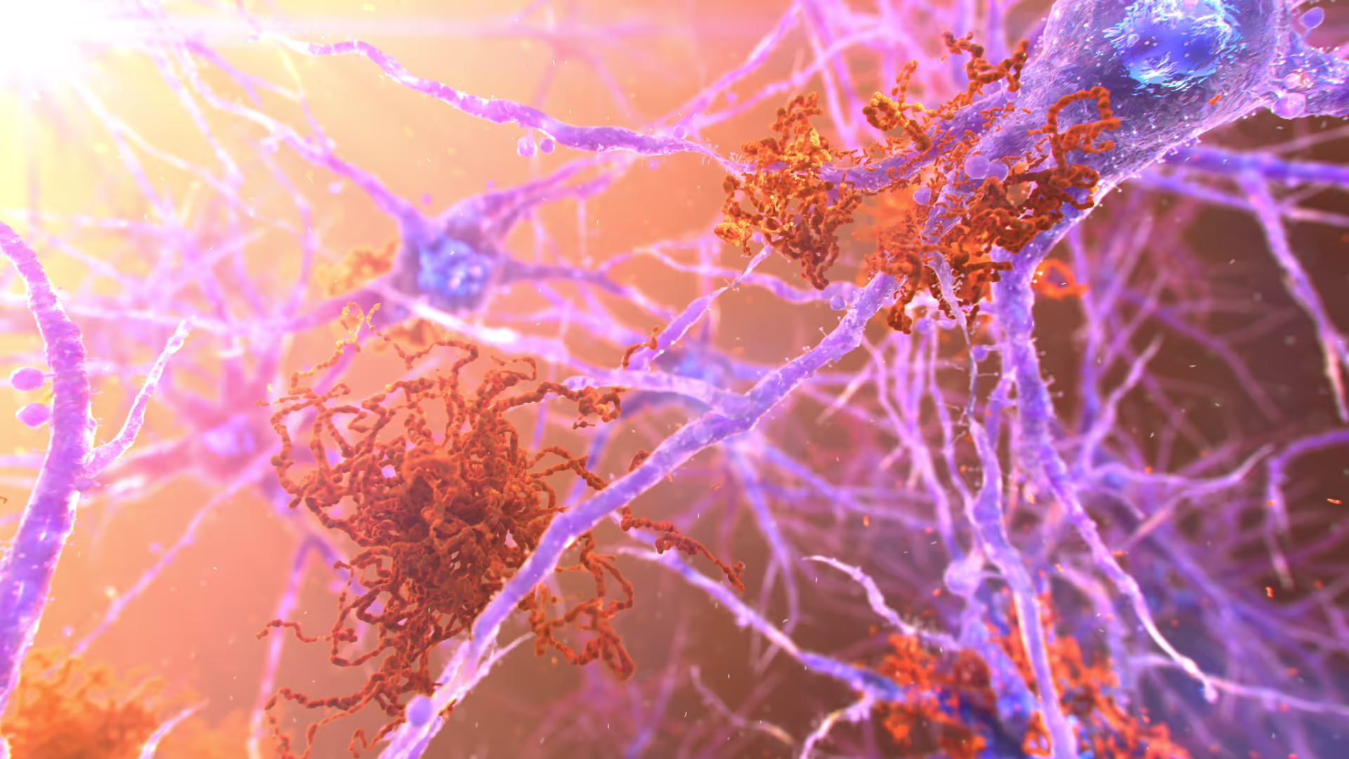
Claire Durrant (UK Dementia Research Institute) went on to discuss how they are using the brain tissue to understand Alzheimer’s and other diseases that display dementia symptoms. Her team was able to trace amyloid-beta outside of the neurones as well as Tau protein inside the neuron.
By mapping the specific brain regions the samples came from, they measured concentrations of amyloid-beta and Tau. Interestingly, higher concentrations were found in the temporal lobe and frontal cortex, even in a broad range of patients who had likely not had an Alzheimer's disease diagnosis. This type of mapping is crucial for early detection and for testing treatments in human models first.
Once again the hope is that testing experimental medicine candidates in living brain tissue before they enter clinical trials, increases the chance of success.
Insights into Glioma Research
Lastly Paul Brennan returned to give an insight into Glioma research. Historically, the focus has been cutting out the tumour.
Now, the focus is shifting to the "security guards" of the brain – the myeloid cells (white blood cells).
The research observed how these cells responded to the spread of tumour cells. In some cases, the white blood cells targeted and killed the cancer cells. In others, a strange interaction occurred where the immune cell left the tumour cell completely unharmed. Understanding why this happens is the next big hurdle in preventing tumour spread.
Take away thoughts
Reflecting on the evening (at the side of a hockey pitch) with a breast surgeon friend, she made an excellent point: breast cancer is so widely studied partly because tissue is readily available. If this initiative can expand to other centres, the potential for Neuroscience research is limitless.
- Human models first: Testing treatments in living human tissue may give more accurate results than animal models allow.
- Optimism for a cure: Claire Durrant was genuinely hopeful that this line of research opens real possibilities for an Alzheimer's disease cure.
- Bespoke collaboration: The evening was a perfect example of what happens when surgeons and scientists collaborate.
I think I share Claire’s optimism for the future.
Thank you to Paul Brennan (consultant neurosurgeon), Claire Durrant (UK Dementia Research Institute) and Sam Booker (Simons Initiative for the Developing Brain) for really fascinating talks. And to Edinburgh Neuroscience for organising.
If you'd like to see how we visualise neuroscience, check out our Neurology Expertise page.
References:
- Liharska, L.E., Park, Y.J., Ziafat, K. et al. A study of gene expression in the living human brain. Mol Psychiatry (2025). https://doi.org/10.1038/s41380-025-03163-1
- Kopell BH, Kaji DA, Liharska LE, Vornholt E, Lund A, Hashemi A, et al. (2025) A study of RNA splicing and protein expression in the living human brain. PLoS One 20(10): e0332651. https://doi.org/10.1371/journal.pone.0332651https://journals.plos.org/plosone/article?id=10.1371/journal.pone.0332651
- Rais, M., Binder, D. K., Razak, K. A., & Ethell, I. M. (2018). Sensory Processing Phenotypes in Fragile X Syndrome. ASN Neuro, 10(1). https://doi.org/10.1177/1759091418801092


.svg)
